

 +1-855-573-2663
 support@secondopinions.com
 May 29, 2024

The most common type of diagnostic “quality control” is a patient-initiated second opinion1. In one study, 38% of patients sought a second opinion because they had doubts about the diagnosis or treatment and 19% sought a second opinion because they were dissatisfied with the level of communication2.
What if AI became a helper in identifying signs of chronic conditions before the patients have symptoms? what if one asks for a diagnosis for one ailment but AI highlights opportunistic findings that are related to other chronic conditions, he was not aware of?
The vast volume of digital data or “big data” in the form of images generated by an aging population, with an ever-increasing demand for imaging, amassed by radiology departments, provides ample opportunity for AI application and has allowed radiology to become a service line leader of AI in the medical field. The screening and detection capabilities of AI make it a valuable tool as the medical field moves towards early identification and intervention, especially as it relates to chronic diseases.
The field of medicine today benefits from a vast collection of data, algorithms, and analytics. AI can sift through all this data at record speed, identify patterns, and provide insight crucial in making informed decisions.
If you think that machines make decisions without oversight, think again.
AI platforms for medical purposes must pass certain standards of precision to be certified.
Furthermore, all insights provided by AI must be approved by a qualified physician.
If you are skeptical of a second opinion rendered by AI technology, consider it’s another tool in the toolbox of the radiologist during the diagnostic evaluation of the exam.
Think of an AI rendered second opinion as indicating and measuring findings for the radiologist to pay attention, a kind of another pair of glasses for the radiologist, not as a replacement for it.
Some researchers have proposed that when an AI-driven diagnosis contradicts the initial diagnosis of a physician, another physician should be sought for yet another opinion. This would allow patients to “trust but verify,” accepting an AI driven diagnosis when it supports initial findings, yet seeking another opinion where it conflicts with the physician’s initial findings3.
Let’s turn to how AI has the potential to help prevent one the deadliest forms of chronic diseases.
Almost half of all adults in the U.S. have at least one form of cardiovascular disease (CVD), making it hard to find someone that hasn’t been affected by the disease in one way or another4.
The U.S. spends $225 billion on CVD every year, and yet it remains the leading cause of death in the US and around the world. CVD is deemed a “silent killer” because so many people are not aware they have it, often showing its first symptom as a major cardiac event, like a stroke or heart attack. In these cases, the healthcare system is reactive rather than proactive, with patients only visiting their doctor when a symptom or issue arises5.
Proactive efforts can include, for example, obtaining a coronary artery calcium (CAC) scoring CT scan, a dedicated imaging study requiring added cost, time and radiation exposure. CAC scores are considered the most reliable predictor for a patient’s future risk of cardiovascular events. However, much of the same information about coronary artery calcium is present on ALL non contrast chest CTs performed for any clinical indication. However, radiologists typically do not evaluate CAC levels on standard chest CT scans if they haven’t been requested to report them.
Today, new artificial intelligence (AI) solutions can assess CAC levels and empower radiologists and cardiologists with the data they need to intercept some forms of heart disease, like CAD, before it is too late. AI algorithms have a remarkable ability to serve as an excellent tool for enhancing personalized healthcare and boosting prognosis. Deep learning (DL) and machine learning (ML) have significantly transformed the field of detecting and forecasting disorders6.
In the utopic world, when radiologists proactively measure CAC levels during routine chest CT scans, they ensure comprehensive reporting that includes crucial information about coronary artery calcium. By integrating these findings into the report with detailed measurements and recommendations for further clinical action, radiologists empower referring physicians to make informed decisions regarding patient care. This proactive approach enhances the likelihood of follow-up and treatment plans, ultimately contributing to improved patient outcomes.
Unfortunately, we are not there yet. Radiologists focus on the clinical indication and while they do their best to issue a complete report, often times chronic findings are not commented on and that is where AI comes in to support the radiologist.
AI may assist physicians by providing them with data-driven insights and predictive analytics, enabling them to make more informed decisions. [6]
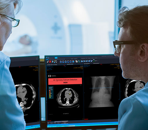
Nanox AI’s cardiac solution is an artificial intelligence software to evaluate calcified plaques in the coronary arteries on non-ECG gated chest, which may present risk for coronary artery disease.
Leveraging the use of routine and existing CT scans, Nanox.AI Cardiac solution integrates seamlessly within a radiologist’s workflow, empowering them with clear, easy-to-interpret visual identifications of CAC levels. Using AI to maximize this already obtained imaging data, can be instrumental to evaluate calcified plaques in the coronary arteries, which present a risk for coronary artery disease, aiming to promote the clinician to suggest the next steps in the care pathway to potentially decrease the risk of a cardiovascular event such as a heart attack or stroke.
The software can proactively identify patients with signs that correlate to risk of a coronary artery disease, promoting appropriate risk assessment, preventative care, and/or follow up treatment so that CVD treatment is proactive and not reactive.
Let’s look at patient care for CAD with and without AI technology; a potential patient case from a US institution.
A patient presented in the Emergency Room with chest and back pain. The ER physician ordered blood tests, an EKG scan, and a CT scan to examine the root cause of the pain.
The CT scan was reported as ‘normal’ apart from a vertebral compression fracture, which could potentially explain the patient’s back pains. This patient’s CAC, present on his Chest CT scan, went unnoticed. Luckily, the Nanox.AI technology for measuring coronary artery calcium on non-contrast chest CTs was deployed at this institution, which subsequently sent the results of high coronary artery calcification levels to the cardiology team. A cardiology consultation was scheduled after their ER visit, and the patient was started on appropriate statin medication, followed by a complete cardiac workup which led to coronary artery stenting.
The unfortunate and often “silent” reality of CVD is commonplace across the world. If AI was implemented, CAC levels could be detected prior to or at the first sign of clinical symptoms of heart disease.
Nanox AI’s cardiac software was run retrospectively on 549 cases in a clinical setting at a Michigan hospital system. The results were reviewed by the lead preventative cardiologists and their team to check the AI results against human findings.
In 49% of the cases, Nanox AI’s software classified CAC as moderate or severe. In 83% of the moderate cases, the physicians agreed with Nanox’s findings. In 92% of the severe cases, the physicians agreed with Nanox’s findings.Overall agreement rate with the software output was 89%.
And most importantly, when Nanox.AI’s cardiac solution detected moderate or severe coronary artery calcium, approximately 65% of those cases had previously been undiagnosed with CAD. In that hospital system, in one year, 3710 patients with medium or high coronary artery calcium were identified without a prior diagnosis or cardiovascular disease.
An ounce of prevention is worth a pound of cure.
Be proactive, take the AI leap, and get a second opinions on your chest CT scan with Nanox.AI today and identify and measure the degree of coronary artery calcium to assess your cardiovascular risk and potentially prevent illness tomorrow.

Our website content is posted for informational purposes only. It is not intended to be used for primary diagnoses-making and should not replace a consultation with a professional health care provider. If you have any health issues or complaints, please consult your primary physician. Healthcare data provided for informational purposes is not an alternative to an in-person physician consultation.
This website is an informative site that aims to offer its users find helpful information regarding a second opinion services that will be suitable for their medical condition. The content provided in this website is not and shall not be taken as expert or professional medical advice for any matter and is not an alternative to an in-person physician consultation. Our services are different from the diagnostic service typically provided by a physician, as the physicians do not have the benefit of information that would be obtained by examining you in person, observing your physical condition, or conducting diagnostic testing to the specifications of the physician. Therefore, the physician may not be aware of facts or information that would affect the physician ́s medical opinion of your condition. In some cases, these facts may be critical to the opinion. USARAD is not responsible for potential errors in opinion resulting from missing, incomplete, poorly translated or illegible records, or poor-quality images